- May 04, 2026
- By Joint Care London
Knee Osteoarthritis: Causes, Symptoms, Stages & Treatment
Knee osteoarthritis is the most common joint condition in the UK — and one of the leading causes of pain, disability and reduced quality of life in adults over 45. Yet despite its prevalence, there remains widespread confusion about what knee osteoarthritis actually is, what causes it, and — crucially — what can be done about it beyond waiting for a joint replacement.
This guide covers everything you need to know: from the anatomy of the knee and how osteoarthritis develops, to the full spectrum of treatment options available today — including the latest injection therapies available at our clinics in London.
What Is Knee Osteoarthritis?
Osteoarthritis (OA) is a degenerative joint disease characterised by the progressive breakdown of articular cartilage — the smooth, rubbery tissue that covers the ends of bones within a joint. In a healthy knee, this cartilage allows the femur (thigh bone), tibia (shin bone), and patella (kneecap) to glide over one another with minimal friction. It also acts as a shock absorber, distributing load across the joint during walking, running, and everyday movement.
In knee osteoarthritis, this cartilage gradually thins, softens, and fragments. As the protective layer wears away, the underlying bone becomes exposed and begins to change in response to increased stress — forming bone spurs (osteophytes), thickening (sclerosis), and sometimes developing fluid-filled cysts. The joint's lining (synovium) becomes inflamed, contributing further to pain and swelling.
The result is a joint that is stiff, painful, and progressively less functional. Knee OA is not simply "wear and tear" — it is a complex biological process involving mechanical, inflammatory, and genetic factors — and it is one that can be meaningfully managed at every stage.
How Common Is Knee Osteoarthritis?
Knee OA affects an estimated 4.7 million people in England alone. Globally, it is the most common form of osteoarthritis and one of the leading causes of disability in older adults. Prevalence rises sharply with age — affecting around 10% of adults over 55 and nearly 50% of those over 75 — but it is increasingly diagnosed in younger adults, particularly those with a history of sporting injury, obesity, or prior knee surgery.
Women are approximately twice as likely as men to develop knee osteoarthritis, particularly after the menopause — a pattern that suggests hormonal factors play a role in cartilage health.
What Causes Knee Osteoarthritis?
Knee osteoarthritis is not caused by a single factor. It develops when cumulative mechanical stress, biological vulnerability, and environmental risk factors combine to overwhelm the knee's natural capacity for repair. Understanding the causes helps explain both why OA develops and how certain treatments work to slow its progression.
Primary Risk Factors
- Age: Cartilage repair mechanisms become less efficient with age. The risk of knee OA increases significantly after 45 and continues to rise with each decade of life.
- Excess body weight: Each kilogram of excess body weight places approximately three to five kilograms of additional force across the knee joint during walking. Obesity is one of the strongest modifiable risk factors for both developing and accelerating knee OA.
- Previous knee injury: Anterior cruciate ligament (ACL) tears, meniscal injuries, fractures around the knee, and patellar dislocations all significantly increase the risk of developing OA in later life — even when well treated at the time.
- Female sex: Women develop knee OA more frequently and more severely than men, particularly after the menopause. Oestrogen appears to have a protective effect on cartilage.
- Genetics: There is a clear hereditary component to osteoarthritis. If a parent or sibling has knee OA, your risk is significantly elevated.
- Joint malalignment: Varus (bow-legged) or valgus (knock-kneed) alignment concentrates load unevenly across the knee compartments, accelerating cartilage wear in the overloaded area.
- Muscle weakness: Weakness in the quadriceps — the muscles at the front of the thigh that help stabilise and cushion the knee — is both a consequence and a driver of knee OA progression.
- Occupational loading: Jobs involving prolonged kneeling, squatting, heavy lifting, or climbing stairs are associated with accelerated cartilage degradation.
- High-impact sport: Elite-level participation in football, rugby, and distance running is associated with increased long-term OA risk, particularly in the presence of prior injury.
Recognising the Symptoms of Knee Osteoarthritis
Knee OA symptoms typically develop gradually over months to years. They are often first noticed during or after physical activity and may fluctuate significantly — with periods of relative calm punctuated by painful flare-ups.
Common Symptoms
- Pain: Usually felt across the front, inner, or outer knee. Pain often worsens with activity — particularly walking on uneven ground, climbing stairs, or rising from a seated position — and eases with rest in the earlier stages. In more advanced disease, pain may persist at rest or disturb sleep.
- Morning stiffness: A characteristic feature of OA is stiffness lasting up to 30 minutes after waking or after prolonged inactivity. This differs from inflammatory arthritis (such as rheumatoid arthritis), where morning stiffness typically lasts over an hour.
- Swelling: The knee may feel puffy or look visibly swollen, particularly after activity. This results from excess synovial fluid (an effusion) produced by the inflamed joint lining.
- Crepitus: A grinding, creaking, or crunching sensation or sound within the knee during movement — caused by roughened joint surfaces moving against one another.
- Reduced range of motion: As OA progresses, the knee may not straighten or bend fully. Many patients notice difficulty squatting, kneeling, or sitting with bent knees for extended periods.
- Bony enlargement: Osteophytes (bone spurs) around the joint may cause a visible or palpable bony prominence around the knee margins.
- Instability or giving way: Muscle weakness and joint surface irregularities can cause the knee to feel unstable or to buckle unexpectedly during weight-bearing.
- Altered gait: Many patients unconsciously adapt their walking pattern to reduce load on the painful knee, which can lead to secondary problems in the hip, lower back, and opposite knee.
How Is Knee Osteoarthritis Diagnosed?
Diagnosis of knee OA combines clinical assessment with imaging. A thorough evaluation is important not only to confirm the diagnosis but to characterise its severity, identify which compartments of the knee are most affected, and rule out other causes of knee pain — including meniscal tears, bursitis, ligament injury, and inflammatory arthritis.
Clinical Examination
A skilled clinician will assess your gait, observe the alignment of the knee, palpate for tenderness and bony enlargement, measure range of motion, test joint stability, and evaluate muscle strength. Specific clinical tests can help identify whether other structures — such as the menisci or ligaments — are contributing to symptoms.
X-Ray
Plain X-ray is the standard first-line imaging investigation for suspected knee OA. It is used to assess four key features:
- Joint space narrowing: Reflecting cartilage loss — the hallmark of OA.
- Osteophyte formation: Bone spurs around the joint margins.
- Subchondral sclerosis: Thickening and hardening of the bone immediately beneath the cartilage.
- Subchondral cysts: Fluid-filled cavities within the bone, indicating advanced disease.
X-ray appearances are graded using the Kellgren-Lawrence (KL) scale from 0 (normal) to 4 (severe bone-on-bone OA). It is important to note that the severity of X-ray changes does not always correlate with the severity of symptoms — some patients with marked radiological OA have relatively mild pain, while others with early changes experience significant disability.
MRI
MRI provides detailed images of cartilage, menisci, ligaments, and bone. It is particularly useful when the diagnosis is uncertain, when symptoms seem out of proportion to X-ray findings, or when a concurrent soft tissue injury (such as a meniscal tear) is suspected and may require separate management.
Ultrasound
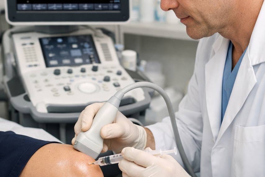
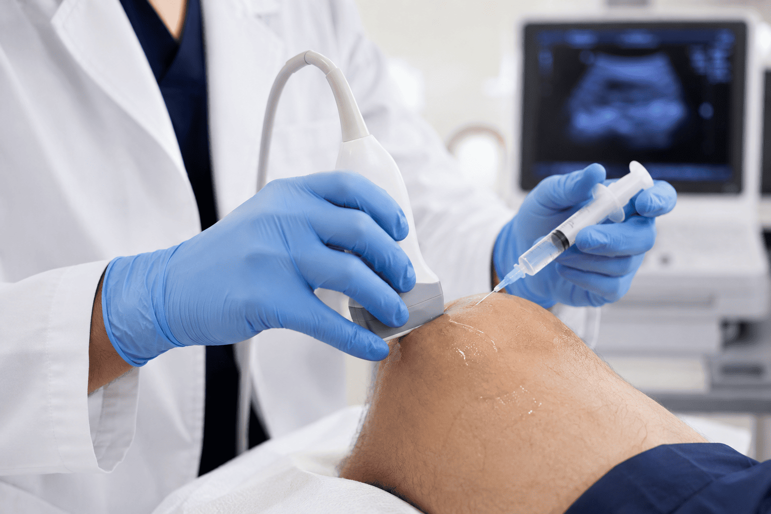
Diagnostic ultrasound can detect joint effusion (excess fluid), synovial thickening, and Baker's cysts (fluid-filled swellings behind the knee). At Joint Care London, ultrasound is also used in real time to guide all knee injections, ensuring precise, accurate medication delivery.
Stages of Knee Osteoarthritis
Understanding the stage of knee OA helps guide treatment decisions. The Kellgren-Lawrence grading system is the most widely used classification:
- Grade 0 — Normal: No radiological features of OA. Symptoms, if present, are likely due to other pathology.
- Grade 1 — Doubtful: Possible minor osteophytes. Uncertain significance. Symptoms may be early or intermittent.
- Grade 2 — Mild: Definite osteophytes with possible joint space narrowing. Symptoms often present with activity but not at rest.
- Grade 3 — Moderate: Multiple osteophytes, definite joint space narrowing, some sclerosis, possible deformity. Symptoms are more persistent and may impact daily activities.
- Grade 4 — Severe: Large osteophytes, marked joint space narrowing, severe sclerosis, definite deformity. Pain at rest and significant functional limitation. Surgical review usually warranted.
Most non-surgical treatments — including injection therapies — are most effective in grades 2 and 3, where meaningful cartilage and joint space remain.
Treatment Options for Knee Osteoarthritis
Knee osteoarthritis cannot currently be cured — cartilage does not fully regenerate. However, a wide range of evidence-based treatments can significantly reduce pain, restore function, slow progression, and delay or potentially avoid the need for surgery. Treatment is most effective when tailored to the individual, taking into account the severity of OA, the patient's symptoms, lifestyle, activity goals, and overall health.
1. Lifestyle Modification and Self-Management
For most patients, lifestyle changes form the essential foundation of knee OA management. These interventions are the most evidence-based of all treatments for OA and carry no side effect profile.
- Weight management: In overweight patients, losing 10% of body weight has been shown to reduce knee pain by up to 50% and slow cartilage loss. Even modest weight loss produces meaningful benefits. This is the single most impactful thing many patients can do.
- Exercise: Counterintuitively, movement is medicine for knee OA. Regular, appropriate exercise maintains muscle strength, preserves range of motion, stimulates cartilage nutrition, and reduces pain. The key is choosing the right type of exercise.
- Activity modification: Reducing or replacing high-impact activities (running on hard surfaces, heavy squatting) with low-impact alternatives (swimming, cycling, walking on even terrain, water aerobics) reduces joint loading while maintaining fitness and function.
- Footwear: Flat, cushioned shoes with good shock absorption reduce forces transmitted to the knee. Lateral wedge insoles may help patients with medial (inner) compartment OA.
- Walking aids: A cane used in the opposite hand to the affected knee can reduce joint load by up to 25% and reduce pain during longer walks.
- Heat and cold therapy: Heat packs relax stiff joints and muscles; ice packs reduce swelling and inflammation during acute flare-ups.
2. Physiotherapy and Exercise Rehabilitation
A structured physiotherapy programme is one of the most effective long-term interventions for knee OA. The primary goal is strengthening the muscles that support and stabilise the knee — particularly the quadriceps, hamstrings, and hip abductors — to reduce the mechanical load placed on the joint surface itself.
Physiotherapy for knee OA typically includes:
- Targeted strengthening exercises (e.g., straight leg raises, wall sits, step-ups)
- Range of motion exercises to maintain joint mobility
- Neuromuscular training to improve balance and proprioception
- Manual therapy techniques to improve joint mechanics
- Hydrotherapy (aquatic physiotherapy) for patients who find land-based exercise too painful
- Education on pacing, activity planning, and joint protection strategies
A commitment of 8–12 weeks of regular physiotherapy is typically required before meaningful benefits are established. Crucially, exercise therapy does not wear the joint out further — in fact, moderate loading stimulates cartilage metabolism and is essential for long-term joint health.
3. Oral Medication
Oral analgesics and anti-inflammatory medications are commonly used for short-term pain management during flare-ups or while other treatments take effect.
- Paracetamol: First-line for mild pain. Safe for most patients when taken at recommended doses, but evidence for its effectiveness in OA is relatively modest.
- NSAIDs (e.g., ibuprofen, naproxen): More effective than paracetamol for OA pain and inflammation, but carry gastrointestinal, cardiovascular, and renal risks with prolonged use. Not suitable for all patients.
- Topical NSAIDs (e.g., diclofenac gel): Applied directly to the skin over the knee, topical formulations provide local anti-inflammatory effects with significantly lower systemic side effects. Recommended as a first-line option alongside or before oral NSAIDs.
- Opioids: Not recommended for long-term OA management due to their unfavourable risk-benefit profile for chronic musculoskeletal pain.
- Supplements: Glucosamine and chondroitin have a large consumer following, but current evidence for their efficacy in knee OA is inconsistent. They are not routinely recommended in UK clinical guidelines, though they are generally safe to try.
4. Injection Therapies for Knee Osteoarthritis
When lifestyle measures and physiotherapy provide insufficient relief, or when pain is severe enough to prevent engagement with rehabilitation, targeted injection therapies can provide meaningful and lasting symptom control. At Joint Care London, all knee injections are performed under real-time ultrasound guidance by experienced musculoskeletal doctors — ensuring precise, accurate placement and optimal outcomes.
Corticosteroid (Steroid) Injection
Corticosteroid injections deliver a concentrated anti-inflammatory directly into the knee joint. They work by suppressing the inflammatory cascade within the synovium, rapidly reducing pain and swelling. Steroid injections are among the most widely used and well-evidenced treatments for knee OA pain, particularly during acute flare-ups or when significant joint effusion is present.
What to expect: Most patients notice improvement within 3–7 days. Relief typically lasts 4–16 weeks, though individual responses vary considerably. Injections are generally limited to 2–3 per year; frequent repeat injections may accelerate cartilage breakdown over time.
Best suited to: Patients with moderate to severe pain, significant swelling, or acute inflammatory flare-ups. Also useful as a short-term bridge to allow engagement with physiotherapy or while waiting for another treatment to take effect.
Hyaluronic Acid (Viscosupplementation) Injection
Hyaluronic acid (HA) is a naturally occurring substance found in healthy synovial fluid. In an osteoarthritic knee, synovial fluid becomes degraded — thinner, less viscous, and less effective as a lubricant and shock absorber. Viscosupplementation involves injecting high-molecular-weight hyaluronic acid directly into the knee joint to restore these properties.
Unlike steroids, which target inflammation, HA works by improving the mechanical and biological environment of the joint. It provides lubrication, reduces the transmission of mechanical stress to the cartilage surface, and may have mild anti-inflammatory and cartilage-protective effects.
What to expect: Benefits develop more gradually than with steroid injections, typically over 2–4 weeks, but tend to last longer — with relief often sustained for 6–12 months or more in good responders. A single-injection formulation of high-molecular-weight HA is offered at Joint Care London.
Best suited to: Patients with mild to moderate OA who want longer-lasting relief, those who have not responded well to steroids, or those who wish to minimise the use of anti-inflammatory medication.
Platelet-Rich Plasma (PRP) Injection
PRP is prepared by drawing a small sample of the patient's own blood, centrifuging it to concentrate the platelets, and injecting the resulting plasma — rich in growth factors and signalling proteins — into the knee joint. These growth factors are thought to modulate inflammation, promote cartilage cell activity, and support tissue repair.
PRP has attracted significant interest as a potential disease-modifying treatment, rather than purely symptomatic. Evidence is growing, particularly for mild to moderate OA, with a number of studies showing benefits in pain and function comparable or superior to hyaluronic acid.
What to expect: Some patients experience a temporary increase in discomfort for a few days after the injection. Improvement typically emerges over 4–6 weeks, with benefits potentially lasting 6–12 months. A course of 1–3 injections is commonly recommended.
Best suited to: Younger patients with early to moderate OA, those seeking a biologically-based approach, or patients who have not had sustained benefit from other injections.
Read our dedicated article on PRP injections: what they are, how they work, and what to expect.
Arthrosamid
Arthrosamid is a non-degradable polyacrylamide hydrogel — a relatively new and innovative option for knee osteoarthritis. Unlike conventional injections that are absorbed by the body over weeks or months, Arthrosamid integrates with the synovial membrane (the joint's inner lining), providing a persistent, long-term cushioning and pain-relieving effect from a single injection.
Clinical evidence, including the multicentre TACIT trial, demonstrates sustained improvements in pain and function over 2–3 years. At Joint Care London, our own clinical data shows that approximately 15–50% of patients experience very significant improvement following Arthrosamid injection.
Best suited to: Patients with confirmed mild to moderate knee OA (KL grade 2–3) who have had insufficient or short-lived benefit from steroid or hyaluronic acid injections, and who wish to delay or avoid knee replacement surgery.
For a detailed breakdown of Arthrosamid, including how it compares to other injection options, read our dedicated guide: Arthrosamid for Knee Osteoarthritis: Evidence, Outcomes and How It Compares to Other Treatments.
5. Surgical Options
Surgery is generally considered when pain is severe and constant, quality of life is significantly impaired, and conservative and injection-based treatments have been exhausted. Surgical options for knee OA include:
Arthroscopic Surgery
Keyhole surgery to wash out the joint (lavage), remove loose cartilage fragments, or address concurrent pathology such as a torn meniscus. Evidence for arthroscopy as a treatment for knee OA itself is limited, and it is no longer routinely recommended for OA in the absence of specific mechanical symptoms or confirmed meniscal pathology.
Osteotomy
A procedure to realign the knee by removing or adding a wedge of bone above or below the joint. Osteotomy is considered in younger, active patients with unicompartmental OA and significant malalignment, aiming to redistribute load away from the most affected compartment. It can delay the need for joint replacement by many years.
Unicompartmental Knee Replacement (UKR)
When OA is confined predominantly to one compartment of the knee (most commonly the inner, medial compartment), a partial knee replacement may be appropriate. UKR replaces only the damaged compartment, preserving the healthy areas of the knee and the cruciate ligaments. Recovery tends to be faster than with total knee replacement, and results in a more natural-feeling knee in appropriately selected patients.
Total Knee Replacement (TKR)
Total knee replacement is the definitive surgical treatment for advanced, end-stage knee OA affecting multiple compartments. The damaged joint surfaces of the femur, tibia, and patella are replaced with metal and plastic implants. TKR is one of the most performed and successful elective surgical procedures in the UK, with excellent long-term outcomes for the right patient. However, it involves major surgery with a recovery period of 3–6 months and carries risks including infection, blood clots, stiffness, and implant-related complications. The average implant lifespan is 15–20 years.
Living Well with Knee Osteoarthritis
A diagnosis of knee osteoarthritis does not mean an inevitable decline into disability. With the right combination of self-management, appropriate treatment, and — where needed — specialist input, the majority of people with knee OA lead active, fulfilling lives for many years.
The following principles have the strongest evidence base for long-term outcomes:
- Stay active: The worst thing for an arthritic knee is inactivity. Regular, low-impact exercise maintains muscle support, preserves range of motion, and reduces pain over time.
- Manage your weight: Even small reductions in body weight translate into meaningful improvements in knee pain and function.
- Engage with physiotherapy: The benefits of supervised exercise rehabilitation are cumulative and long-lasting — but require consistent effort over weeks and months.
- Seek help early: Early intervention — before cartilage loss becomes severe — gives the widest range of treatment options and the best long-term outcomes.
- Don't wait for a perfect solution: Knee OA management works best as a combination of approaches tailored over time, not a single silver-bullet treatment.
Frequently Asked Questions: Knee Osteoarthritis
Is knee osteoarthritis the same as arthritis?
Osteoarthritis is the most common form of arthritis, but "arthritis" is a broader term covering over 100 different joint conditions — including rheumatoid arthritis, gout, and psoriatic arthritis. Knee OA is specifically a degenerative condition caused by cartilage breakdown, distinct from inflammatory arthritis, which is driven by the immune system attacking the joints.
Can knee osteoarthritis get better on its own?
Knee OA does not reverse without intervention — cartilage does not regenerate significantly once lost. However, symptoms can fluctuate considerably. Flare-ups often resolve with rest and appropriate management. With the right treatment and lifestyle changes, many people experience sustained periods of good symptom control and stable disease.
What is the best exercise for knee osteoarthritis?
Low-impact aerobic exercise — swimming, cycling, walking, and water aerobics — combined with targeted quadriceps and hip strengthening exercises provides the greatest benefit with least joint stress. High-impact activities such as running or jumping should be modified but not necessarily abandoned entirely, depending on the individual and the severity of OA.
How do I know if I need a knee replacement?
Knee replacement is generally considered when pain is severe, constant (including at rest and at night), significantly impairs daily activities and quality of life, and has not responded to a sustained course of conservative and injection-based management. The decision should be made collaboratively with an orthopaedic surgeon following appropriate imaging and clinical assessment.
Are knee injections safe?
Knee injections performed by experienced clinicians under ultrasound guidance are very safe. Risks include temporary post-injection discomfort, infection (very rare when performed under sterile conditions), and — with repeated steroid use — potential effects on the surrounding tissues. Our doctors will discuss the specific risks and benefits of each injection type during your consultation.
How quickly will I feel the benefit from a knee injection?
Steroid injections typically provide noticeable relief within 3–7 days. Hyaluronic acid and PRP injections tend to work more gradually — over 2–4 weeks — but may provide longer-lasting benefit. Arthrosamid typically shows meaningful improvement at around the four-week mark, with continued gradual improvement over the following months.
Can I have a knee injection if I am on blood thinners?
This depends on the specific medication and your clinical circumstances. Some blood thinners require temporary adjustment before a joint injection; others do not. This will be reviewed in full during your consultation at Joint Care London before any procedure is undertaken.
Knee Osteoarthritis Assessment and Treatment at Joint Care London
At Joint Care London, we provide rapid-access assessment and injection treatment for knee osteoarthritis at our clinics in Notting Hill and Golders Green. We offer the full range of knee injection therapies — steroid, hyaluronic acid, PRP, and Arthrosamid — all performed under real-time ultrasound guidance by experienced musculoskeletal doctors.
We understand that knee pain affects not just your mobility, but your sleep, your work, your independence, and your enjoyment of life. Our goal is to provide fast access to the right treatment for you — with appointments typically available within days, not weeks, and at a fraction of the cost of a private hospital procedure.
If knee pain is holding you back, contact us today to arrange a consultation. Together, we can help you find the most effective approach for your specific situation and get you back to doing the things that matter.